MRI MACHINE: Cost, Working, Benefits & Risks
An MRI machine is a medical imaging device that uses strong magnetic fields and radio waves to create detailed images of the inside of the body. MRI stands for magnetic resonance imaging. They are commonly used to diagnose and monitor a wide range of medical conditions such as tumors, cancer, heart and vascular diseases, fractures, cartilage and ligament disorders, and spine problems like stenosis, disc prolapse, etc. MRI machine has revolutionized the viewing of structures inside the body, especially the soft tissues of the-
- Brain
- Spine
- Abdomen
- Joints
MRI Machine Price
MRI is an expensive machine, and its price ranges from Rs 1 Crore to Rs 7 Crore in India. The actual price depends on
- Brand
- Tesla (0.5 to 3 Tesla)
- Technology
- Feature (Silent, in-built music, etc.)
- Protocols/ Software
Some of the popular brands for MRI in India are:
- Philips
- GE
- Siemens
- Hitachi
MRI Machine Details in Brief
| Also Known as | Magnetic Resonance Imaging machine |
| Purpose | To visualize the inside of the body especially soft tissues |
| Preparation | Remove any jewellery or other metal objects |
| Technique | It uses strong magnets and radio waves |
| Test Type | Imaging Test |
| Time taken | 30 mins to 1 hour (in most cases) |
| Popular Brands | Philips, GE, Siemens, Hitachi |
| Cost (INR) | Rs 1 crore to Rs 7 crore |

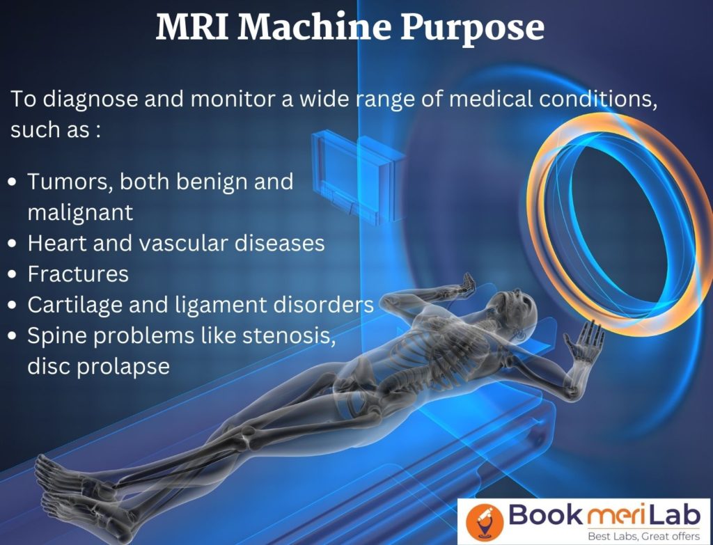
MRI Machine Purpose
The medical Conditions that can be diagnosed & assessed using MRI Machines
- Tumors– It can help distinguish between malignant (cancerous) and benign (non-cancerous) growths. They can also be used to evaluate the extent of cancer spread and to monitor the response to cancer treatment.
- Heart and vascular diseases like coronary artery disease, heart valve disease, abnormal iron deposition in the heart, congenital heart diseases, cardiac tumors, aneurysms.
- Orthopedic Diseases like
- Spine Problems– Degenerative disc diseases like prolapse and herniation, spinal stenosis, facet joint issues, etc, disorders of cartilage, tendons, ligaments, or bones
- Fractures
- Tearing or rupture of ligaments, tendons, or other soft tissues
- Cartilage disorders
- Joint disorders-osteoarthritis, rheumatoid arthritis, and ligament or tendon injuries.
- Brain-related issues like clots of cerebral vessels, disorders of the eye and inner ear, brain tumors, Multiple sclerosis, strokes, etc.
- To Guide a Biopsy Procedure: An MRI scan can help the doctor locate the area of the body that needs to be biopsied. It can provide important information about the tissue or organ’s size, shape, and location.
- To Guide Surgery: An MRI scan can help the doctor determine the best surgical approach and provide important information about the location and size of the structures that will be affected by the surgery. During the surgery, an MRI machine can help the doctor see exactly what they are doing during the surgery and provide important feedback on the procedure’s progress. For example, an MRI machine can guide a needle during a spinal injection or monitor a surgical procedure’s effects on the brain or spinal cord. After the surgery, an MRI machine can be used to evaluate the procedure’s results and monitor the patient’s recovery. The images can help the doctor assess the effectiveness of the surgery and can provide important information about any complications or side effects.
Preparation
To prepare for an MRI, the patient may be asked to remove any jewellery or other metal objects that could interfere with the images, and they may be asked to wear a hospital gown. The patient will need to lie still on the table during the scan, which typically takes 30 minutes to an hour. The MRI machine is typically noisy, so the patient may be given earplugs or headphones to wear during the scan.
After the scan, the images are analyzed by a radiologist, and the results are provided to the patient’s doctor, who will interpret the images and discuss the findings with the patient.

How Does an MRI Machine Look Like?
- An MRI machine is a hollow cylindrical chamber consisting of a large, tube-shaped magnet, a radio frequency (RF) transmitter, and a receiver.
- The magnet generates a strong magnetic field, and the transmitter and receiver send and receive the radio waves. These waves generate signals of different intensities to view and distinguish the different structures in the body.
- The patient lies on a moveable table inserted into the center of the MRI machine, and the magnet, RF transmitter, and receiver create detailed images of the body’s tissues and organs.
Open MRI
Open MRI machines typically have a flatbed and a wider bore, or opening, than a traditional MRI machine and may also have a shorter tunnel. This allows the patient to have more room and feel less confined during the scan. This is especially helpful for claustrophobic patients.
Standing MRI
A standing MRI machine is a type of MRI machine that allows patients to stand or sit upright during the imaging process. This type of MRI machine is typically used for imaging the spine, joints, and other parts of the body that may be difficult to scan in a traditional lying-down MRI machine.
Different Types of MRI Machines
Some of the most common types of MRI machines include:
High-field MRI machines: These are the most common type of MRI machines and they generate a magnetic field strength of 1.5 T to 3.0 T or more. They are also larger and more expensive than other types of MRI machines.
Low-field MRI machines: It generates a magnetic field strength of 0.2 T to 0.5 T, which is lower than the field strength of high-field MRI machines. Low-field MRI machines are typically smaller and less expensive than high-field MRI machines, but they may not be able to produce images with as much detail.
Open MRI machines: These MRI machines have a more open design, with a wide opening that allows the patient to be less enclosed during the scan. They are typically used for patients who are claustrophobic or who have difficulty lying still during a scan. However, open MRI machines may not be able to produce images with as much detail as high-field or low-field MRI machines.
High-resolution MRI machines: These MRI machines use advanced technology to produce images with higher resolution than other types of MRI machines. They are typically used for specific applications, such as imaging the small bones of the hand or foot, and they may be more expensive than other types of MRI machines.
Benefits of MRI Machines
Some of the key advantages of MRI machines include the following:
High-resolution images: They can produce images with high spatial resolution, showing small structures and details in the body’s tissues and organs.
No ionizing radiation: MRI machines do not use ionizing radiation, which means that they do not carry the same risks as X-ray or CT scans.
Safe for patients with metal implants: Safe for patients with MRI-compatible metal implants, such as pacemakers or metal clips in the brain. Because MRI machines use magnetic fields and radio waves rather than X-rays, so they are less affected by metals than other medical imaging tests.
Multiplanar imaging: MRI machines can create images in multiple planes, producing images of the body’s tissues and organs from different angles. This can provide important information about the shape and orientation of structures in the body.
Functional imaging (fMRI): It produces functional images showing how the body’s tissues and organs function. For example, an MRI scan can show how blood flows through the arteries or how the brain responds to a particular task.
Risks and Limitations of MRI Machines
Some of the potential risks and limitations of MRI machines include the following:
Metal in the Body: Metal implants: MRI is generally safe for people with metal implants, but in some cases, the strong magnetic field used in the scan can cause these devices to move or heat up. This can be dangerous, especially for implants near the heart or brain. Any iron-containing magnetic implant in the body poses fear of displacement in the MRI Machine.
Claustrophobia: Some people may feel claustrophobic or anxious when they are inside the MRI machine. This can make it difficult for them to lie still during the scan, affecting the image quality. For patients who are claustrophobic, there are open MRI machines available that have a more open design, but these machines may not be able to produce images with as much detail.
Noise: MRI machines are typically noisy, which can be loud and unpleasant for some patients, especially those sensitive to noise or who have hearing impairments. Patients may be given earplugs or headphones to wear during the scan to reduce the noise.
Length of the scan: MRI scans typically take 20 minutes to an hour.
Cost and availability: MRI machines are expensive. Thus, this can make it difficult for some patients to access MRI services, especially if they live in rural or underserved areas.
Pregnancy: MRI machines do not use ionizing radiation but produce strong magnetic fields, which may harm the developing baby during pregnancy. For this reason, MRI scans are generally not recommended for pregnant women unless there is a compelling medical reason to have the scan.
Allergies to contrast agents: Some MRI scans require the use of contrast agents, which are substances that are injected into the body to enhance the visibility of certain structures on the images. These contrast agents can cause allergic reactions in some people, and they may not be safe for patients with allergies or previous reactions to contrast agents.
History of MRI Machines
The history of MRI machines dates back to the early 20th century when scientists first discovered the magnetic resonance effect, which is the phenomenon that allows MRI machines to produce detailed images of the inside of the body.
- In the 1920s, scientists in Germany and the United States began to study the magnetic resonance effect and explore its potential applications in medical imaging. In the 1940s and 1950s, scientists developed the first MRI machines, which were large and cumbersome, and were used primarily for research purposes.
- Later, in the 1960s and 1970s, MRI technology continued to advance, and scientists developed new techniques and pulse sequences that allowed MRI machines to produce detailed images of the body’s tissues and organs. In the 1980s, MRI machines became more widely available and were used increasingly for diagnostic purposes.
Some of the key scientists involved in the early history of MRI include:
Felix Bloch and Edward Purcell: In the 1940s, these two physicists independently discovered the phenomenon of NMR and shared the 1952 Nobel Prize in Physics for their work.
Raymond Damadian: In the early 1970s, Damadian, a medical doctor and physicist, developed the first MRI machine, which he called the “Indomitable.”
Paul Lauterbur: In the early 1970s, Lauterbur, a chemist, developed the technique of magnetic resonance imaging (MRI), which allowed for the creation of two-dimensional images of the body using NMR. He was awarded the Nobel Prize in Physiology or Medicine in 2003 for this work.
Peter Mansfield: In the early 1980s, Mansfield, a physicist, developed the technique of magnetic resonance imaging (MRI), which allowed for the creation of detailed, three-dimensional images of the body using NMR. He was awarded the Nobel Prize in Physiology or Medicine in 2003 for this work.
How MRI Machines Work: Basic Principle
The basic principles of MRI machines are based on the principles of nuclear magnetic resonance (NMR), which was first discovered in the 1940s.
- NMR is a physical phenomenon that occurs when the nuclei of atoms are exposed to a strong magnetic field and radio waves. The nuclei of atoms, such as hydrogen atoms, have a property called spin, which causes them to behave like tiny magnets. When they are placed in a strong magnetic field, they align with the field and begin to spin.
- When a radio frequency (RF) pulse is applied to the nuclei, they absorb the RF energy and change their spin state. This causes them to release a small amount of energy in the form of radio waves, which can be detected by the MRI machine’s receiver. By measuring the amount of energy released and the time it takes to release the energy, the MRI machine can create detailed images of the body’s tissues and organs.
- MRI machines are typically large, tube-shaped machines that generate a strong magnetic field using superconducting magnets. The patient lies on a moveable table that is inserted into the center of the MRI machine, and the machine’s RF transmitter and receiver create detailed images of the body’s tissues and organs.
- The magnetic field strength of an MRI machine is typically measured in units of tesla (T), and the strength of the field can range from 0.5 Tesla to 3.0 Tesla or more. The stronger the magnetic field, the more detailed the images will be, but higher field strengths can also increase the MRI machine’s cost and the scan’s length.
- The MRI machine can also use different pulse sequences, or combinations of RF pulses, to create images with different contrast and resolution and of different cross sections. For example, a T2-weighted pulse sequence can create images that highlight fluid-filled structures, such as the cerebrospinal fluid in the spinal cord, while a T1-weighted pulse sequence can create images that highlight bone and other dense tissues.
Advise to Patients
Before the MRI scan, your doctors will review your medical history. They will also conduct a physical examination to determine if an MRI scan is appropriate for your specific needs. This is a good time to ask any questions about the MRI scan and discuss any concerns you may have.
FAQs
An MRI (magnetic resonance imaging) machine is a medical imaging device that uses strong magnetic fields and radio waves to produce detailed images of the inside of the body.An MRI (magnetic resonance imaging) machine is a medical imaging device that uses strong magnetic fields and radio waves to produce detailed images of the inside of the body.An MRI (magnetic resonance imaging) machine is a medical imaging device that uses strong magnetic fields and radio waves to produce detailed images of the inside of the body.
An MRI machine uses a powerful magnetic field to align the nuclei of atoms in the body, and radio waves to create a rotating magnetic field. This causes the nuclei to produce a faint signal, which is detected by the MRI machine and used to create detailed images of the body.
MRI scans are generally considered to be safe, as they do not use ionizing radiation. However, there are some risks associated with MRI scans, such as the risk of allergic reactions to contrast agents, or the risk of being affected by the strong magnetic field.
MRI scans are used for a wide range of medical purposes, including imaging of the brain, spine, and joints, as well as for detecting abnormalities in the body, such as tumors, injuries, or diseases.
Most people can get an MRI scan, but there are some exceptions. For example, people with certain medical implants, such as pacemakers or certain types of metal implants, may not be able to undergo an MRI scan due to the risk of interference with the device. It’s important to discuss your medical history with your doctor before undergoing an MRI scan
Dr Garima Saroj
Dr Garima Saroj is a dentist from Manipal college of dental sciences. She has previously worked at ESIC dental college and hospital Delhi and Dentecare multispecialty dental clinic Gurugram. She has a passion for writing for patient education and awareness.